
Page 25 - IJEEE-2022-Vol18-ISSUE-1
P. 25
Received: 13 October 2021 Revised: 17 November 2021 Accepted: 18 November 2021
DOI: 10.37917/ijeee.18.1.3
Vol. 18| Issue 1| June 2022
? Open Access
Iraqi Journal for Electrical and Electronic Engineering
Original Article
Brain MRI Images Segmentation Based on U-Net
Architecture
Assalah Zaki Atiyah*, Khawla Hussein Ali
Department of Computer Science, College of Education for Pure Sciences, University of Basrah, Basrah, Iraq
Correspondence
* Assalah Zaki Atiyah
College of Education for Pure Sciences,
University of Basrah, Basrah, Iraq
Email: pgs2179@uobasrah.edu.iq
khawla.ali@uobasrah.edu.iq
Abstract
Brain tumors are collections of abnormal tissues within the brain. The regular function of the brain may be affected as it grows
within the region of the skull. Brain tumors are critical for improving treatment options and patient survival rates to prevent
and treat them. The diagnosis of cancer utilizing manual approaches for numerous magnetic resonance imaging (MRI) images
is the most complex and time-consuming task. Brain tumor segmentation must be carried out automatically. A proposed strategy
for brain tumor segmentation is developed in this paper. For this purpose, images are segmented based on region-based and
edge-based. Brain tumor segmentation 2020 (BraTS2020) dataset is utilized in this study. A comparative analysis of the
segmentation of images using the edge-based and region-based approach with U-Net with ResNet50 encoder, architecture is
performed. The edge-based segmentation model performed better in all performance metrics compared to the region-based
segmentation model and the edge-based model achieved the dice loss score of 0. 008768, IoU score of 0. 7542, f1 score of 0.
9870, the accuracy of 0. 9935, the precision of 0. 9852, recall of 0. 9888, and specificity of 0. 9951.
KEYWORDS: Brain Tumor, Convolution Neural Network (CNN), Edge Segmentation, Region Segmentation, U-Net.
I. INTRODUCTION Segmenting medical images is the initial step in
diagnosing, planning, and investigating brain tumor disease.
Brain tumors are abnormal brain tissue collections. A Currently, tumor segmentation is done manually by a
very rigid skull protects the brain. In such a confined space, radiologist, which takes a long time. It may take several
growth might cause issues. Brain tumors are classified as hours for a single patient to perform the task, and radiologists
benign or malignant. A benign or malignant tumor may need to concentrate for a long time.
expand the skull tumor. Tumors may be identified by their
origin. The majority of brain cancer cells diseases are Gliomas are a form of tumor that needs therapy as soon
primary tumors of the brain. The most commonly diagnosed as it is identified in a patient, hence rapid segmentation is
cells in the brain moved from different parts of the body are required. Auto-tumor-segmentation is superior to manual
secondary (metastatic) brain tumors [1]. Magnetic resonance tumor-segmentation in speed and accuracy. It will also
imaging is a widely used tool for diagnosing brain cancers. minimize the period between diagnostic testing and therapy,
There are several magnetic resonance sequences, each allowing clinicians to focus on the patient's health and design
focusing on a different kind of normal or abnormal tissue [2]. a treatment plan.
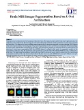
Native T1-weighted (T1), post-contrast T1-weighted (T1ce),
T2-weighted (T2), and T2 fluid-attenuated inversion recovery Deep neural networks have recently attracted researchers
(T2-FLAIR) MR modalities were used in this study. Figure 1 due to their great performance and accuracy in image
shows all these modalities. segmentation[3]. A CNN can recognize and infer
characteristics from images. Many research has utilized
Fig. 1: Four Different Modalities of MRI CNN to segment brain tumors on MRI images. This research
proposes a method to segment brain tumors. Images are
segmented using edge-based and region-based methods. The
brain tumors are segmented using U-Net with ResNet50
encoders.
This paper presents methods for the segmentation of brain
tumors using region-based and edge-based approaches. This
This is an open access article under the terms of the Creative Commons Attribution License, which permits use, distribution and
reproduction in any medium, provided the original work is properly cited.
© 2021 The Authors. Iraqi Journal for Electrical and Electronic Engineering by College of Engineering, University of Basrah.
https://doi.org/10.37917/ijeee.18.1.3 https://www.ijeee.edu.iq 21